*Human Brain Mapping and Cortical Imaging Laboratory, International Doctoral School in Biomedical Sciences & Engineering, Aalborg University, Denmark +Dept. of Mathematical Modelling, Tech Univ of Denmark, Lyngby, Denmark
Background
Neuroimaging of human pain has advanced rapidly. Topographic mapping of EEG/ERP activation and tomographic registration of PET/fMRI responses requires systematic examination. Talairach coordinates in neuroimaging now provide a universal anatomical standard [1]. To facilitate visualization and data organization, a 3D brain model of VRML/Talairach has been devised [2]. The information registration and retrieval of brain activation and elucidation of function can also be performed by hypertext in this computer 3D model. Using pain of headache and heart-attack as the data basis, this work illustrates the use of this 3D Brain Model in examination of brain loci and organization associated with human pain.
Methods
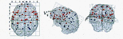
We compiled activated foci from published PET studies. Cluster headache (n= 7) was induced by infusion of nitroglycerin [3] and angina (n= 12) with dobutamine [4]. Colour/shape coding is used to denote both increase and decrease of rCBF. The sites of significant SPM activation are depicted by single dots and clusters of similar dots representing either headache (red/square: rCBF increase and blue/round: rCBF decrease) or heart-attack (red/diamond: rCBF increase and blue/cross: rCBF decrease).
Results
For the Talairach sites during pain of headache and heart-attack, a large disperse of PET activation can be observed in the figures. Interestingly, the PET increase sites of headache (red square) and heart-attack (red diamond) were more in the anterior forebrain. However, it is surprising that the PET decreases in headache (blue round) and heart-attack (blue cross) are confined largely in the posterior brain. There is no discernible pattern of laterality effect. From these figures, there is hardly any overlapping of PET activation sites between the headache and heart-attack conditions.
|
|
Conclusion
Our analysis of the 3D brain model during pain of headache and heart-attack serves as
an illustration of how the computerized Talairach representation can be used in neuroimaging of human pain.
The 3D Brain can be used for house keeping the brain activation in pain research [5].
A large scale statistical analysis is anticipated using this system.
References
1. Talairach J & Tournoux P. Co-Planar Stereotaxic Atlas of the Human Brain, Suggart: Thieme Verlag, 1988.
2. Nielsen F.Å, Hansen L.K. Neuroimage, 7(4):S782, 1998.
3. Hseich JC, et al. Pain, 67(l):59-68, 1996.
4. Rosen SD, et al. Lancet, 344:147-50, 1994.
5. Chen ACN, http://www.smi.auc.dk/HBMCI, 1999. (ac@smi.auc.dk)
Acknowledgement
This project has been funded by the Danish Research Council as well as the Danish National Research Foundation.